

7 October 2026 Punjab Khabarnama Bureau : Diagnosing lesions located deep within the neck can be challenging because of the region’s complex anatomy and the presence of critical structures such as major blood vessels, nerves, bones and air-filled spaces. New research suggests that CT-guided core needle biopsy can provide a reliable and minimally invasive option when conventional ultrasound-guided biopsy is difficult or impossible.
A recent study published in European Radiology evaluated 122 CT-guided core needle biopsies performed in 118 patients with lesions in ultrasound-inaccessible suprahyoid deep neck spaces. Researchers found a diagnostic yield of 93.4%, indicating that the technique was able to provide adequate diagnostic information in the overwhelming majority of procedures.
The study also found that CT-guided biopsy had an 89.5% concordance rate with surgical pathology among cases in which surgical comparison was available. Minor procedure-related complications occurred in 4.1% of cases, supporting the procedure’s overall safety when performed in appropriate clinical settings.
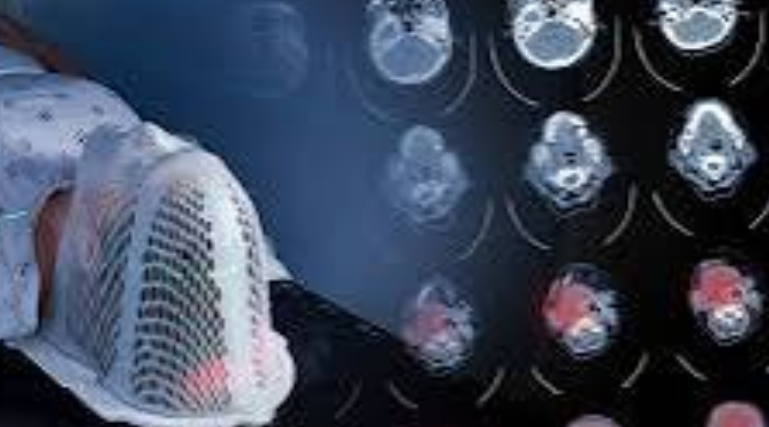
Deep neck lesions can be particularly difficult to evaluate because ultrasound waves may be obstructed or degraded by bones and air-containing structures. This can make it difficult for clinicians to clearly visualize the target and safely guide a needle into the lesion. CT imaging, by contrast, can provide detailed cross-sectional views of the anatomy and help physicians plan a needle trajectory around important structures.
Earlier research involving 204 patients similarly demonstrated the potential of CT-guided core needle biopsy for deep suprahyoid head and neck lesions. In that study, all biopsy specimens were considered adequate for histological diagnosis, while overall diagnostic accuracy reached 91.2%. The sensitivity was 89.2% and specificity was 97.8%.
The researchers noted that CT guidance is particularly useful for lesions involving areas such as the skull base, parapharyngeal space, parotid region and other deep anatomical compartments that may not be readily accessible with ultrasound.
The procedure involves carefully planning the needle path using CT imaging before tissue is collected. Depending on the location of the lesion, physicians can use different approaches to reach the target while attempting to avoid blood vessels, nerves and other vulnerable structures.
A coaxial biopsy technique can also allow multiple tissue samples to be obtained through a single access route. Obtaining a sufficient amount of tissue is important because many deep neck lesions require detailed histopathological evaluation to distinguish between benign and malignant conditions and to determine the appropriate treatment strategy.
Researchers caution, however, that CT-guided biopsy is not guaranteed to produce a correct diagnosis in every case. Lesions with poorly defined margins can be more difficult to target accurately, increasing the risk of sampling error. In such situations, detailed pre-procedural imaging, including MRI when appropriate, may help physicians better identify the boundaries and most informative portion of a lesion.
The findings are particularly relevant for patients with suspected head and neck tumors, where obtaining tissue confirmation can be essential before treatment. A minimally invasive biopsy may help physicians avoid a more invasive surgical procedure when a safe and diagnostically adequate needle pathway is available.
The latest research supports the selective use of CT guidance as a complementary technique rather than a replacement for ultrasound-guided biopsy. Ultrasound remains useful for many superficial neck lesions, while CT can offer an important alternative when lesions are deep or obscured by anatomical structures.
Experts emphasize that the procedure requires substantial radiological expertise because the head and neck contain numerous closely packed critical structures. Careful imaging review, selection of the safest needle trajectory and appropriate patient selection remain important factors in achieving accurate results.
Overall, the evidence indicates that CT-guided core needle biopsy can provide a high diagnostic yield for challenging deep neck lesions, particularly when ultrasound-guided tissue sampling is not feasible. Further research and broader clinical experience could help refine patient selection, procedural techniques and strategies for improving diagnostic accuracy in complex head and neck cases.