

1 October 2026 Punjab Khabarnama Bureau : Researchers at the Indian Institute of Technology Hyderabad have developed a novel bioink designed to closely reproduce the biological and mechanical environment of lung tissue, potentially opening new possibilities for lung tissue engineering, regenerative medicine, disease modelling and drug testing.
The research focuses on a major challenge in 3D bioprinting: creating artificial tissue that does not merely resemble the physical structure of an organ but also provides the biological signals needed to guide cells and regulate immune responses. The IIT Hyderabad team developed a lung-derived extracellular matrix bioink that can support stem cell development while encouraging an anti-inflammatory response in immune cells.
The study, published in the journal Biomaterials, was led by researchers Soham Ghosh and Professor Falguni Pati of IIT Hyderabad’s Department of Biomedical Engineering. The researchers investigated decellularized lung matrix bioinks prepared using an osmotic decellularization strategy. The findings showed that preserving important components of the lung’s extracellular matrix could improve both the biological performance and printing characteristics of the resulting material.
The extracellular matrix, or ECM, is the complex network of proteins and other molecules surrounding cells. It provides structural support while also sending biochemical signals that influence cell growth, differentiation and function. Replicating these signals is particularly important in tissue engineering because synthetic materials can provide structural support without necessarily reproducing the biological cues present in natural tissue.
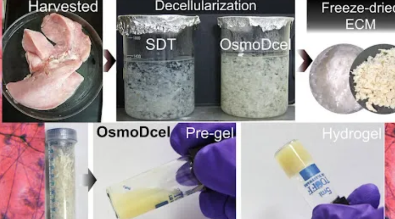
To address this problem, the researchers developed an osmotic decellularization approach designed to preserve key components of the lung matrix. The process uses changes in salt concentration to break down and remove cells while retaining important extracellular components, including sulfated glycosaminoglycans and basement membrane proteins.
These preserved components are significant because they contribute to the biochemical environment of native lung tissue. Conventional detergent-based decellularization methods can remove some of these molecules along with cellular material, potentially reducing the ability of the resulting scaffold to reproduce the natural tissue environment.
The researchers converted the decellularized lung matrix into a printable bioink and examined how it behaved during extrusion-based 3D bioprinting. A major challenge in bioprinting soft tissues is finding the right balance between biological softness and structural stability. Materials need to be soft enough to resemble the target tissue but sufficiently stable to retain their printed shape.
The team used thixotropy testing and extrusion-related measurements to determine how the bioink responded to mechanical stress during printing and how quickly it recovered after leaving the printer nozzle. This allowed the researchers to identify printing conditions that supported structural recovery and multilayer fabrication.
The resulting hydrogels retained mechanical characteristics within the range associated with lung tissue while remaining compatible with cells. This combination is important because lung tissue is relatively soft and compliant compared with many other organs.
The biological results were particularly significant. When mesenchymal stem cells were placed on the surface of the printed lung-derived scaffolds, they showed markers associated with epithelial development. The researchers observed expression of epithelial proteins and surfactant-associated genes, indicating that the lung-derived environment could influence stem cell fate.
The response varied depending on how the cells were positioned within the scaffold. Cells located within the material developed characteristics associated with an interstitial myogenic phenotype, demonstrating that the three-dimensional microenvironment could influence cell behaviour in a spatially dependent manner.
The bioink also demonstrated an immunomodulatory effect. Macrophages, a type of immune cell involved in inflammation and tissue repair, showed an anti-inflammatory profile when cultured within sulfated-glycosaminoglycan-rich lung matrix scaffolds. This response remained evident even when the cells were exposed to inflammatory stimulation.
The finding is important for regenerative medicine because excessive or prolonged inflammation can interfere with tissue repair. A successful tissue-engineering scaffold therefore needs to provide structural and biological support while avoiding an immune environment that could damage newly developing tissue.
The researchers say the work demonstrates that preserving specific components of native lung extracellular matrix can help create a bioink that combines printability, appropriate mechanical behaviour, cell-instructive properties and immunomodulatory activity.
The potential applications extend beyond future tissue replacement. Such printed lung models could provide researchers with new platforms for studying respiratory diseases and testing medicines under laboratory conditions. They could also help scientists investigate how lung cells interact with their surrounding matrix and immune cells.
However, the technology remains at the research stage. The study provides important in-vitro evidence, but a laboratory scaffold is not equivalent to a fully functioning human lung. Future research will need to examine how these constructs behave in living systems, including their long-term integration, vascularisation, immune responses and ability to maintain appropriate lung functions.
The development is part of IIT Hyderabad’s broader research efforts in bioengineering, 3D bioprinting and regenerative medicine. The institute’s Biomedical Engineering research programmes include the development of tissue and organ constructs, novel bioinks and in-vitro models for disease research and drug testing.
By combining native lung-derived biological signals with 3D printing technology, the new approach offers a way to build tissue-engineering scaffolds that are designed to do more than provide physical support. Instead, they can potentially guide cell behaviour and help create a more regenerative microenvironment.
The researchers’ findings highlight the growing role of extracellular-matrix-based materials in regenerative medicine and demonstrate how preserving the biochemical complexity of natural tissues could improve the performance of future bioprinted organs and tissue models.